From AI Diagnostics to 3D-Printed Crowns: How Technology is Reshaping Modern Dentistry
Published 20 min read
Introduction: Why Modern Dentistry Is Changing So Quickly
Dentistry has always relied on sharp eyes, steady hands, and years of clinical experience. But something significant is shifting. Digital tools are moving the profession away from purely manual, judgment-heavy workflows and toward faster, data-driven approaches that can catch problems earlier and deliver results more consistently. Artificial intelligence is now reading X-rays, intraoral scanners are replacing goopy impression trays, and 3D printers are producing crowns right in the dental office. This article explores how AI diagnostics, digital treatment planning, and 3D-printed restorations are reshaping what happens from the moment a patient sits in the chair to the moment they walk out with a finished restoration.
Throughout this piece, you'll get a clear look at the most important themes surrounding dental technology today — accuracy, efficiency, patient outcomes, and yes, the real limits of automation. One thing worth stating upfront: none of these tools replace the dentist. AI can flag a suspicious shadow on a radiograph, but a trained clinician still interprets it, confirms the diagnosis, and decides on treatment. The technology is powerful, but the dentist is still very much in charge. With that framing in mind, let's dig into what's actually happening in modern dental practices.
What AI Diagnostics Means in Dentistry
AI diagnostics in dentistry refers to software that analyzes dental radiographs, intraoral images, cone beam CT scans, and patient records to help detect disease and support clinical decision-making. These systems use machine learning algorithms — often deep learning models trained on thousands or millions of annotated dental images — to recognize patterns that indicate pathology. Think of it as giving the dentist a highly trained second set of eyes that never gets tired and can process images in seconds. The software doesn't replace the dentist's judgment; it gives them more information to work with before they make a call.
The range of conditions these tools can help identify is broader than most patients realize. AI systems are currently being used to flag early caries, signs of periodontal bone loss, periapical pathology, oral lesions that may indicate cancer, sinus disease visible on dental imaging, and temporomandibular joint disorders. Many published reviews report that AI-assisted analysis improves both diagnostic accuracy and the speed at which findings are identified. For a busy practice seeing dozens of patients a day, that kind of efficiency gain isn't trivial — it can meaningfully change how care is delivered.
That said, it's important to understand what AI is and isn't doing here. These systems are decision support tools, not autonomous diagnosticians. They surface findings, highlight areas of concern, and help clinicians avoid missing something on a crowded radiograph. Multiple authoritative reviews in the dental literature are consistent on this point: human oversight isn't just recommended, it's essential. The dentist reviews the AI's output, applies their clinical knowledge, considers the patient's full history, and makes the final call. AI handles the data processing; the dentist handles the patient.
How AI Improves Dental Image Analysis and Early Detection
Machine learning and deep learning models are particularly well-suited to image analysis, which makes them a natural fit for dentistry. These algorithms can scan a full-mouth series of X-rays, a panoramic radiograph, or a CBCT scan and flag abnormalities in a fraction of the time it would take a clinician to review them manually. The models are trained to recognize subtle visual patterns — slight changes in radiodensity, early interproximal caries that barely show through enamel, or the early stages of bone resorption around an implant — that might be easy to miss during a high-volume clinic day.
Early detection is where AI arguably delivers its biggest clinical benefit. Studies have shown that AI-assisted caries detection can identify lesions at earlier stages than traditional manual review alone, giving patients a better shot at conservative treatment rather than more invasive intervention later. AI tools have also demonstrated improvements in sensitivity and specificity for detecting periapical pathology and periodontal bone loss. Perhaps just as importantly, these tools can reduce interobserver variability — the frustrating reality that two equally trained dentists looking at the same X-ray don't always agree on what they see. AI helps bring more consistency to that process.
Early detection isn't just a clinical win — it has real implications for treatment timing, patient prognosis, and how efficiently a practice runs. Catching a small cavity before it reaches the pulp means a simpler, cheaper restoration and a better outcome for the patient. Identifying early bone loss around a tooth or implant before it becomes severe means more treatment options and a longer-lasting result. And when AI flags findings quickly and consistently, it helps practices move through diagnostic workflows faster without sacrificing thoroughness. That combination of better outcomes and better efficiency is a compelling reason why adoption of these tools is accelerating.
Evidence, Accuracy, and Current Clinical Performance of AI Tools
The published evidence supporting AI performance in dental imaging has grown substantially over the past several years. Systematic reviews and meta-analyses have reported strong pooled diagnostic metrics for AI-assisted detection of caries, periapical lesions, and periodontal bone loss, with many studies showing accuracy comparable to or better than experienced clinicians under controlled conditions. Interpretation time improvements have also been documented, with AI-assisted review reducing the time needed to analyze complex imaging datasets. These aren't minor incremental gains — in some studies, the performance differences are clinically meaningful.
However, the evidence also reveals an important gap that the dental research community has been honest about. Many AI systems perform impressively at the tooth level — identifying a specific lesion on a specific tooth — but struggle more with full-patient diagnostic charting, where the system needs to integrate findings across an entire dentition and patient history to generate a comprehensive assessment. This distinction matters because real clinical care is rarely about a single tooth in isolation. It involves understanding the patient's overall oral health status, risk factors, and treatment priorities. Some current AI systems handle this integration better than others, and it's an active area of development.
Perhaps the most consistent message across authoritative dental AI research is this: the field needs more external validation, prospective clinical studies, and real-world testing before fully autonomous AI use becomes routine. Most studies to date have been conducted under controlled conditions using curated datasets, which don't always reflect the messiness of everyday clinical practice. Images in the real world vary in quality, patient populations differ, and edge cases abound. The tools are impressive, but the scientific community is appropriately cautious about overstating their readiness for unsupervised clinical deployment. That caution is a feature, not a flaw.
Common Applications Across Dentistry Today
AI is showing up across nearly every corner of modern dental practice. In restorative dentistry, it assists with caries detection on bitewing and periapical radiographs. In periodontics, it helps assess bone levels and identify signs of disease progression. Oral surgeons and implant specialists use AI-assisted planning tools to evaluate bone volume, density, and anatomy before placing implants. Orthodontists are using AI for cephalometric landmark identification and treatment planning support. And in oral medicine and pathology, AI screening tools are being explored to help identify suspicious lesions that warrant further evaluation.
Beyond direct diagnosis, AI is also being used to support risk stratification — helping clinicians identify which patients are at higher risk for disease progression and need more frequent monitoring. Automated follow-up tracking, prognosis assessment, and workflow automation are other areas where these tools add value. A practice using AI-assisted charting, for example, can document findings more quickly and consistently, reducing the chance that something gets overlooked during a busy afternoon of back-to-back appointments. These workflow improvements might sound unglamorous, but they have a real impact on care quality and practice sustainability.
Authoritative sources in the dental literature consistently frame AI as a tool for improving consistency and helping clinicians manage increasingly large volumes of data. As practices grow and patient panels expand, the volume of imaging data that needs to be reviewed and interpreted grows with them. AI doesn't eliminate the need for clinical expertise — it helps clinicians apply that expertise more efficiently and with better information. Think of it less as replacing the dentist's brain and more as giving that brain a faster, more organized filing system that never misplaces anything.
Digital Workflows: From Scans to Treatment Planning
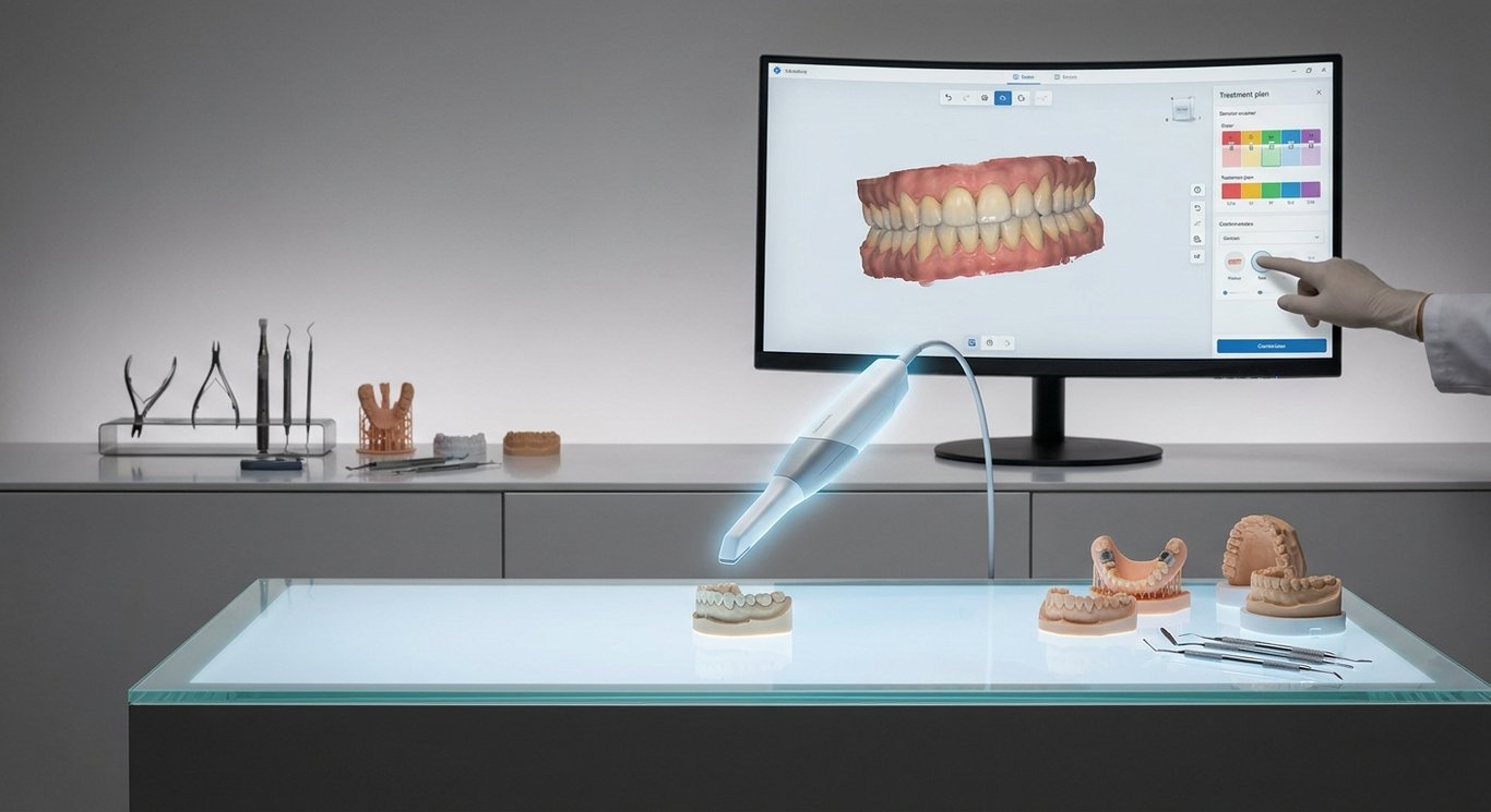
Modern dental technology doesn't work in isolation — it works best when individual tools are connected into a seamless digital workflow. Intraoral scanners capture precise 3D models of a patient's teeth and soft tissue without the need for traditional impressions. CBCT imaging provides detailed volumetric data on bone structure, root anatomy, and surrounding tissue. Digital patient records tie everything together, creating a comprehensive data picture that feeds directly into diagnosis, treatment design, and restoration fabrication. When these systems communicate with each other, the result is a faster, more accurate, and more coordinated care process.
Digital treatment planning reduces the number of manual steps between diagnosis and delivery of care. In implant dentistry, for example, a clinician can use CBCT data combined with an intraoral scan to virtually plan implant placement, assess bone anatomy, and design a surgical guide — all before the patient returns for their procedure. In restorative dentistry, digital design tools allow the clinician to create a crown or bridge virtually, check margins and occlusion on screen, and send the file directly to a milling machine or 3D printer. Communication with dental labs is also improved, since digital files can be shared instantly and reviewed collaboratively without physical models changing hands.
Patients benefit from digital workflows in ways that are immediately tangible. Gone are the days of gagging on impression material while waiting for it to set. Intraoral scanners are faster, more comfortable, and often more accurate. Patients can also see their own teeth in 3D on a screen, which makes it much easier for clinicians to explain what's happening and why treatment is needed. Appointments become more predictable, wait times for restorations can shrink dramatically, and the overall experience feels more modern and professional. For many patients, that shift in experience is just as meaningful as the clinical improvements happening behind the scenes.
3D-Printed Crowns, Bridges, and Other Restorations
3D printing — also called additive manufacturing — has made significant inroads into dentistry over the past decade. In a dental context, it refers to the process of building physical objects layer by layer from a digital design file. Dentists and labs are now using 3D printers to fabricate crowns, bridges, dentures, nightguards, surgical guides for implant placement, orthodontic models, and even temporary restorations. The process starts with a digital scan or design file and ends with a physical object that can be placed directly in the patient's mouth or used as part of a larger treatment process.
The appeal of 3D printing in dentistry comes down to a few key advantages: speed, customization, and repeatability. A crown that once required a physical impression, a trip to an off-site lab, and a two-week wait can now be designed and printed in a fraction of the time. Because the process is driven entirely by digital files, each restoration is precisely tailored to the patient's anatomy. And because the same file can be reprinted if needed, consistency is built into the process in a way that traditional manual fabrication can't always guarantee. For practices that want to offer same-day or next-day restorations, 3D printing is a game-changer.
Of course, patients and clinicians alike want to know: are 3D-printed crowns actually as good as traditional ones? The honest answer is that it depends. Material selection matters enormously — different resins and ceramics have different strength, wear, and esthetic properties, and not all printed materials are suitable for every clinical situation. Fit and marginal accuracy have improved significantly as printer technology has advanced, and several studies have shown printed restorations to be clinically acceptable for many applications. That said, case selection remains important. High-stress posterior restorations, for example, require careful material and design choices to perform reliably over time. The technology is genuinely impressive, but it's not a one-size-fits-all solution.
Benefits for Dentists and Patients
For clinicians, the benefits of adopting AI and digital workflows are both clinical and operational. AI-assisted diagnostics can improve detection rates and reduce the risk of missed findings, which has direct implications for patient outcomes and liability. Digital documentation is faster, more consistent, and easier to retrieve than paper-based records. Streamlined workflows mean less time spent on administrative tasks and more time focused on actual patient care. For dentists who are managing busy practices, these efficiency gains can be significant — not just in terms of time saved, but in terms of reducing the cognitive load of managing large volumes of patient data.
Patients, meanwhile, experience a noticeably different kind of dental visit. Faster turnaround times for restorations mean fewer temporary crowns and fewer return appointments. Intraoral scanners and digital planning tools make procedures more comfortable and more predictable. AI-assisted early detection means problems are caught sooner, often when they're smaller and easier to treat. And the ability to show patients their own imaging data in real time — explained clearly with visual aids — helps them understand their oral health in a way that builds trust and encourages better compliance with treatment recommendations. That's a better experience for everyone involved.
There's also a business case worth acknowledging. Digital dentistry can improve chairside efficiency, which means more patients seen per day without sacrificing quality of care. Better diagnostic tools can improve case acceptance, because patients who can see a problem clearly on a screen are more likely to agree to treatment. And reduced remake rates for restorations — thanks to more accurate digital design and fabrication — mean lower material costs and fewer unhappy patients returning with ill-fitting crowns. For practices willing to invest in the technology and training, the return on investment can be substantial over time.
Limitations, Risks, and Ethical Concerns
AI in dentistry is genuinely exciting, but it comes with real limitations that deserve honest discussion. One of the most significant is the issue of biased training data. AI models learn from the datasets they're trained on, and if those datasets don't reflect the full diversity of patients, imaging equipment, and clinical conditions that exist in the real world, the model's performance in practice may be worse than its performance in controlled testing. Annotation errors in training data — where the images used to teach the algorithm were incorrectly labeled — can also degrade model quality in ways that aren't always obvious. And in atypical or complex cases, AI systems can struggle in ways that an experienced clinician would not.
Privacy and legal responsibility are also serious concerns. Dental AI systems process sensitive patient data, including imaging files and health records. How that data is stored, who has access to it, and how it's protected from breaches are questions that every practice needs to answer before adopting any AI tool. On the liability side, the question of who is responsible when an AI-assisted diagnosis turns out to be wrong is still being worked out legally in many jurisdictions. If an AI system misses a lesion and the dentist accepts that finding without independent review, the legal and ethical responsibility still falls on the clinician. The technology doesn't transfer accountability.
Authoritative reviews in the dental AI literature are remarkably consistent on what's needed to address these concerns: rigorous validation on diverse, real-world datasets; transparency about how models make their decisions; mandatory clinician supervision of AI outputs; and secure, privacy-compliant data handling practices. These aren't just nice-to-haves — they're prerequisites for responsible deployment of AI in a clinical setting. The technology has enormous potential, but realizing that potential responsibly requires the field to hold it to a high standard. That's not pessimism; it's good science.

FDA-Cleared AI and Regulatory Adoption in Dental Imaging
One important signal of how seriously the dental AI space is being taken is the growing number of FDA-cleared products already on the market. The U.S. Food and Drug Administration has cleared several dental AI tools for clinical use, covering tasks such as caries detection on radiographs, periodontal bone level assessment, cephalometric landmark identification for orthodontic analysis, automated dental charting, and anatomical segmentation of CBCT data. FDA clearance means these products have gone through a regulatory review process demonstrating that they are safe and effective for their intended use — a meaningful bar that separates them from unvalidated software tools.
For dentists and patients, FDA clearance provides an additional layer of confidence that a product has been evaluated against established standards. But it's worth being clear about what clearance does and doesn't mean. It doesn't mean the tool is infallible, and it doesn't mean clinical judgment can be set aside. A cleared AI tool for caries detection is still a decision support tool — the dentist reviews its output and makes the final diagnostic call. Regulatory clearance is a floor, not a ceiling. It tells you the tool meets a minimum standard of safety and performance; it doesn't tell you the tool is right in every case. Clinicians who understand this distinction will use these tools most effectively.
How Dental Practices Can Adopt These Technologies
For practices thinking about integrating AI and digital tools, the adoption process works best when it's approached systematically rather than all at once. The first step is identifying which tools are compatible with existing imaging systems, practice management software, and clinical workflows. Not all AI platforms integrate seamlessly with all imaging hardware, and compatibility problems can create more friction than they solve. Once compatible tools are identified, the next priority is staff training — not just on how to use the software, but on how to interpret its outputs and communicate findings to patients. A tool that the team doesn't understand or trust won't deliver its full value.
Evaluating return on investment is a legitimate and important part of the adoption decision. Practices can track time savings in diagnostic workflows, reductions in missed findings, patient satisfaction scores, and remake rates for digital restorations compared to traditional methods. These metrics give a concrete picture of whether the investment is paying off. It's also worth tracking less obvious benefits, like reduced staff stress from more streamlined workflows, or improved case acceptance rates when patients can see their imaging data explained clearly. ROI in dentistry isn't always just about chair time — it's about the overall health of the practice.
That said, implementation barriers are real and shouldn't be minimized. The upfront cost of intraoral scanners, 3D printers, and AI software subscriptions can be substantial, particularly for smaller practices. There's a learning curve involved in shifting to digital workflows, and some team members will adapt faster than others. Interoperability between different software platforms remains an ongoing challenge in the industry. And ongoing quality control — regularly reviewing whether AI outputs are accurate and clinically appropriate — requires time and attention that busy practices may struggle to carve out. None of these barriers are insurmountable, but going in with realistic expectations makes the transition much smoother.
FAQ: Common Questions About AI Diagnostics and 3D-Printed Crowns in Dentistry
Is AI in dentistry accurate enough to trust? Studies and systematic reviews consistently show strong diagnostic performance for AI tools in dental imaging, particularly for caries detection, periapical pathology, and periodontal assessment. In many controlled studies, AI performance is comparable to or better than experienced clinicians. However, "accurate enough to trust" doesn't mean "accurate enough to use without oversight." Dentists still need to review AI findings, apply their clinical knowledge, and make the final diagnostic decision. Think of AI as a highly capable assistant — one that's worth listening to, but not one you'd put in charge without supervision.
Can AI replace dentists? No — and this is one of the clearest points of agreement across authoritative sources in the dental AI literature. AI is designed and validated as a decision support tool, not as a substitute for clinical expertise. Dentistry involves far more than reading images. It requires building patient relationships, exercising clinical judgment in complex situations, performing manual procedures with precision, and making treatment decisions that account for the whole person, not just the data on a screen. AI can make dentists more efficient and more accurate, but it cannot replicate the full scope of what a trained clinician does. The dentist isn't going anywhere.
Are 3D-printed crowns as good as traditional crowns? The honest answer is: it depends on the specifics. 3D-printed crowns have improved significantly in terms of fit accuracy, material strength, and esthetic quality. For many clinical situations, they are a highly effective and efficient option. However, success depends on several factors — the quality of the digital scan, the design of the restoration, the material selected, and whether the case is appropriate for printed fabrication. High-stress posterior restorations require careful material selection to ensure durability. When these factors are managed well, printed crowns can perform excellently. When they're not, problems can arise just as they would with any restoration method.
What dental problems can AI detect? AI tools in dentistry are currently being used to help detect a wide range of conditions. These include interproximal and occlusal caries, periodontal bone loss, periapical pathology, root fractures, calculus deposits, and anatomical variations visible on CBCT. AI is also being used to support oral cancer screening by flagging suspicious lesions in intraoral images, to assist with implant planning by analyzing bone volume and density, and to identify pathological findings in the sinuses or temporomandibular joints that are incidentally visible on dental imaging. The list of applications continues to grow as the technology matures and more validated tools reach the market.
Is digital dentistry safe and regulated? Yes — with important caveats. Many digital dental tools, including AI diagnostic software and imaging systems, are subject to regulatory oversight. In the United States, FDA-cleared AI tools have undergone review for safety and effectiveness before reaching the market. However, not every software product used in dental practices has been cleared, and clinicians should verify the regulatory status of any tool they adopt. Data privacy is also a critical concern — patient imaging data must be stored and transmitted in compliance with HIPAA and other applicable regulations. And as with any clinical tool, proper validation, staff training, and ongoing quality review are essential to safe and effective use.
Conclusion: What the Future of Modern Dentistry Looks Like
The picture that emerges from all of this is one of genuine, meaningful progress. AI is improving how dental diseases are detected and how early clinicians can intervene. Digital workflows are making care faster, more precise, and more comfortable for patients. And 3D printing is fundamentally changing how restorations are designed and fabricated — moving production from off-site labs to chairside, same-day delivery in many cases. These aren't distant promises or experimental technologies; they're tools that are already in use in forward-thinking practices around the world. The transformation of modern dentistry is well underway, and the pace of change is only accelerating.
If you're a dental professional, now is a great time to explore how the technologies covered in this guide — from AI diagnostics to 3D-printed crowns — can improve your diagnostic accuracy, streamline your workflows, and deliver better experiences for your patients. If you're a patient, understanding these advances can help you ask better questions and make more informed decisions about your own care. Either way, the most important thing to keep in mind is that all of this technology works best when it's guided by skilled, attentive clinicians who put patient outcomes first. The future of dentistry is digital — but it's still deeply, fundamentally human.